By Yin Nwe Ko
Knee pain is a common complaint among adults and is most often associated with general wear and tear from daily activities like walking, bending, standing, and lifting. Athletes who run or play sports that involve jumping or quick pivoting are also more likely to experience knee pain and problems. But whether an individual’s knee pain is caused by ageing or injury, it can be a nuisance and even debilitating in some circumstances.
The knee is a vulnerable joint that bears great stress from everyday activities, such as lifting and kneeling, and from high-impact activities, such as jogging and aerobics.
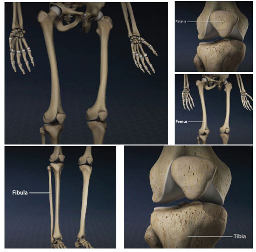
The knee is formed by the following parts:
Tibia: This is the shin bone or larger bone of the lower leg.
Femur: This is the thighbone or upper leg bone.
Patella: This is the kneecap.
Each bone end is covered with a layer of cartilage that absorbs shock and protects the knee. The knee is 2 long leg bones held together by muscles, ligaments, and tendons. There are 2 groups of muscles involved in the knee, including the quadriceps muscles (located on the front of the thighs), which straighten the legs, and the hamstring muscles (located on the back of the thighs), which bend the leg at the knee. tendons are tough cords of tissue that connect muscles to bones. Ligaments are elastic bands of tissue that connect bone to bone. Some ligaments on the knee provide stability and protection of the joints, while other ligaments limit forward and backward movement of the tibia (shin bone). Many knee problems are a result of the ageing process and continual wear and stress on the knee joint (such as arthritis). Other knee problems are a result of an injury or a sudden movement that strains the knee. Common knee problems include the following:
Sprained or strained knee ligaments and/or muscles
A sprained or strained knee ligament or muscle is usually caused by a blow to the knee or a sudden twist of the knee. Symptoms often include pain, swelling, and difficulty in walking.
Torn cartilage
Trauma to the knee can tear the menisci (pads of connective tissue that act as shock absorbers and also enhance stability). Cartilage tears can often occur with sprains. Treatment may involve wearing a brace during an activity to protect the knee from further injury. Surgery may be needed to repair the tear.
Tendonitis
Inflammation of the tendons may result from overuse of a tendon during certain activities such as running, jumping, or cycling. Tendonitis of the patellar tendon is called jumper’s knee. This often occurs with sports, such as basketball, where the force of hitting the ground after a jump strains the tendon.
Arthritis
Osteoarthritis is the most common type of arthritis that affects the knee. Osteoarthritis is a degenerative process where the cartilage in the joint gradually wears away. It often affects middle-aged and older people. Osteoarthritis may be caused by excess stress on the joint such as repeated injury or being overweight. Rheumatoid arthritis can also affect the knees by causing the joint to become inflamed and by destroying the knee cartilage. Rheumatoid arthritis often affects persons at an earlier age than osteoarthritis.
In addition to a complete medical history and physical exam, other tests for knee problems may include:
X-ray
This test uses invisible electromagnetic energy beams to make images of internal tissues, bones, and organs onto film.
Magnetic Resonance Imaging (MRI)
This test uses large magnets, radiofrequency, and a computer to make detailed images of organs and structures within the body; can often determine damage or disease in a surrounding ligament or muscle.
Computed Tomography Scan (CT Scan)
This test uses X-rays and computer technology to make horizontal, or axial, images (often called slices) of the body. A CT scan shows detailed images of any part of the body, including the bones, muscles, fat, and organs. CT scans are more detailed than general X-rays.
Arthroscopy
A minimally-invasive diagnostic and treatment procedure used for conditions of a joint. This procedure uses a small, lighted, optic tube (arthroscope), which is inserted into the joint through a small incision in the joint. Images of the inside of the joint are projected onto a screen; used to evaluate any degenerative or arthritic changes in the joint; detect bone diseases and tumours; to determine the cause of bone pain and inflammation.
Radionuclide bone scan
A nuclear imaging technique uses a very small amount of radioactive material, injected into the patient’s bloodstream to be detected by a scanner. This test shows blood flow to the bone and cell activity within the bone.
When you walk on level ground, the force on your knees is equivalent to about 1.5 times your body weight. It’s no wonder they play up.
What can go wrong?
The knee is a complex joint and we give it a lot of grief over our lifetime by walking, lifting, kneeling, and high-impact activities, such as running, jumping, and sports. Blows to the knee and sports where you are often twisting it are a common cause of injury, including sprained ligaments and muscles, torn cartilage, tendonitis (sometimes caused by excessive running, jumping, or cycling), bursitis (from excessive kneeling), and, as you get older, osteoarthritis.
What if it’s arthritis?
In osteoarthritis, the cartilage in the joint wears away. The main symptoms are pain, stiffness, and difficulty moving the joint. Having arthritis in your knee also makes you more prone to ligament injuries, as the muscles around the joint can become weak. It’s important to exercise to combat pain, improve leg strength and balance, Car and maintain flexibility. Acupuncture may help with osteoarthritis pain.
How do you treat a knee injury?
First off, you’ll need RICE. This means Resting your knee for a day or two, applying Ice—such as a bag of frozen peas wrapped in a tea towel— for up to 20 minutes every two or three hours to bring down the swelling, compressing with a bandage, and elevating your injury above the level of your heart. Don’t play sports if you’re in pain, but if you’re itching to get going, try swimming or another low-impact activity, such as tai chi or pilates, once things have settled down.
And if it doesn’t get better?
See your GP and set about getting a proper diagnosis. They might refer you to a physiotherapist—or you can opt to go privately—who can give you exercises to strengthen the muscles around your knee. Some injuries, such as torn ligaments, might need surgery.
What are the best ways to care for my knees?
Prevention is better than cure. Warm up properly before you exercise and consider sports that are easier on your knees. These include swimming, water aerobics, cycling (on the road or a static bike), pilates, brisk walking, or stepping up. A rule of thumb: never do an exercise where your knees stick out beyond your toes when you bend them. You might want to avoid deep squats and running. Wear shoes with good tread, whether you’re exercising or simply out and about.
How do I make my knees stronger?
Focus on exercises that work your hamstrings, quadriceps, glutes, and hip muscles. These will also help to strengthen your knees. There are plenty of exercises to improve knee strength online.
What difference does body weight make?
Being overweight puts extra strain on your knees, raising the risk of injuries and osteoarthritis. Even modest weight loss will help. Combine losing a few kilos with exercises that tone the muscles around the joint, and your knees will look better too.
Reference: www.hopkinsmedicine.org/health/conditions